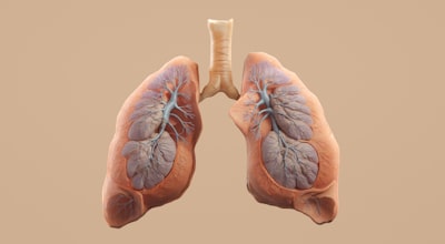
Lung ultrasound (LUS) has become an indispensable bedside tool for assessing pulmonary edema in patients at risk of heart failure or kidney disease. However, its automated analysis faces significant challenges due to speckle noise, imaging artifacts, and operator-dependent acquisition variability. In this context, deep learning offers promising solutions but requires approaches that integrate clinical knowledge to improve accuracy and interpretability. This article explores a LUS video classification framework based on two key components: hierarchy-aware training and anatomy-guided learning, demonstrating how these strategies can overcome the limitations of traditional flat models.
The proposed approach starts from a strong baseline and introduces hierarchical training strategies that organize clinical classes —healthy, B-lines, consolidations, and mixed (B-lines with consolidations)— into a structure that reflects pathological progression. Unlike flat classification where all categories are treated equally, the hierarchy allows the model to learn subordination relationships between classes, improving separation among pathological conditions. For example, distinguishing between isolated B-lines and consolidations becomes more effective when the model first recognizes the presence of pathology and then refines the subtype. This architecture not only increases accuracy but also aligns model behavior with clinical reasoning.
Additionally, a pleural line mask supervision is incorporated, focusing the model's attention on this key anatomical structure in lung ultrasound. By guiding attention toward this region, more localized and anatomically relevant attention patterns are obtained. Experiments on an open dataset of 1,886 videos from 219 patients, evaluated with patient-level five-fold cross-validation, show that pleural mask supervision achieves a mean macro-F1 of 65.7%, outperforming models using only hierarchical training without anatomical guidance. Moreover, transfers to the external COVID-BLUeS dataset demonstrate competitive and parameter-efficient adaptation while preserving pleural-focused attention behavior.
These results have important implications for developing AI-assisted diagnostic systems in clinical settings. The combination of clinically structured objectives with anatomy-guided supervision not only improves performance but also provides interpretability crucial for clinical adoption. Clinicians need to understand why a model classifies a video as pathological; visualizing attention maps that focus on the pleural line builds trust and facilitates integration into workflows.
For health technology companies, this case study underscores the importance of developing custom software that incorporates specific domain knowledge. It is not enough to apply generic computer vision models; software customization to adapt to the particularities of lung ultrasound —such as equipment variability, acquisition techniques, and anatomical definitions— requires a multidisciplinary approach. Here is where companies like Q2BSTUDIO add value, combining software development expertise with understanding of clinical and technical challenges.
Implementing AI solutions in radiology and ultrasound involves handling large volumes of data, often stored in the cloud. Cloud AWS/Azure services provide the scalability needed to train complex models and deploy them in production environments. Additionally, cybersecurity is critical when handling patient data; platforms must comply with regulations like GDPR and HIPAA. Q2BSTUDIO offers cybersecurity solutions that ensure sensitive information protection, integrating security protocols from design.
Another relevant element is the integration of these systems with Business Intelligence (BI) platforms. Analyzing large volumes of LUS videos can generate performance metrics, pathological trends, and early alerts. Using Power BI, hospitals can visualize dashboards correlating ultrasound findings with clinical outcomes, facilitating data-driven decision-making. Furthermore, process automation —from image acquisition to report generation— reduces administrative burden and accelerates diagnosis.
The concept of AI agents also becomes relevant. A LUS classification system could act as an agent that not only classifies but also suggests complementary explorations, alerts about possible acquisition errors, and communicates with other hospital systems. Q2BSTUDIO develops customized AI agents that integrate into clinical workflows, offering intelligent real-time assistance.
In summary, hierarchical and anatomy-guided learning represents a significant advance in automated lung ultrasound analysis. This approach not only improves accuracy and interpretability but also establishes a framework for developing custom software applications that respond to specific clinical needs. Collaboration between research teams and technology companies like Q2BSTUDIO is essential to translate these advances into daily practice, offering robust, secure, and scalable solutions that leverage the latest innovations in AI, cloud, and BI.
The results presented in this study validate the feasibility of combining clinical hierarchies with anatomical supervision. The improvement in macro-F1 and attention localization are indicators that the model learns more meaningful representations. For future implementations, it is recommended to explore multimodal architectures integrating additional clinical data and using reinforcement learning techniques to optimize acquisition sequences. In any case, the key is to develop custom software that captures the complexity of the clinical environment and offers tangible value to healthcare professionals.
In conclusion, the combination of hierarchical learning strategies and anatomy-guided supervision proves to be a practical and robust approach for lung ultrasound video analysis. This work lays the foundation for more interpretable and effective AI systems in diagnosing pulmonary edema, and highlights the importance of developing personalized software that integrates clinical knowledge. Companies like Q2BSTUDIO, with expertise in AI, cloud, cybersecurity and BI, are in a privileged position to drive these innovations in the healthcare sector.